41 diagram of the heart without labels
CBSE Class 10 Science Important Biology Diagrams For Last Minute ... CBSE Class 10 Chemistry Important Reactions. 2. Brain. A human brain is composed of three main parts- the forebrain, the midbrain and the hindbrain. These three parts have specific functions ... Heart: illustrated anatomy - e-Anatomy - IMAIOS This interactive atlas of human heart anatomy is based on medical illustrations and cadaver photography. The user can show or hide the anatomical labels which provide a useful tool to create illustrations perfectly adapted for teaching. Anatomy of the heart: anatomical illustrations and structures, 3D model and photographs of dissection.
Circulatory System Diagram - New Health Advisor There are different types of circulatory system diagrams; some have labels while others don't. The color blue stands for deoxygenated blood while red stands for blood which is oxygenated. Below you'll see diagram specified to the heart, as well as circulatory system diagram of the whole body: How Does the Human Circulatory System Work? 1. Heart
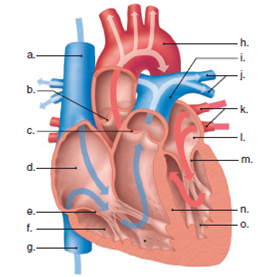
Diagram of the heart without labels
Blank ear diagrams and quizzes: The fastest way to learn - Kenhub Take a moment to look at the ear model labeled above. This shows you all of the structures you've just learned about in the video, labeled on one diagram. Seeing them all together in this way is a great way to learn, since anatomical structures do not exist in isolation. That's why labeling the ear is an effective way to begin your revision. How the Heart Works - The Heart | NHLBI, NIH The heart is an organ about the size of your fist that pumps blood through your body. It is made up of multiple layers of tissue. Your heart is at the center of your circulatory system. This system is a network of blood vessels, such as arteries, veins, and capillaries, that carries blood to and from all areas of your body. Your blood carries ... Draw The Venn Diagram Of A-B : Heart Diagram With Labels â€" Heart ... Heart Diagram With Labels â€" Heart Diagram Without Labels from 5 draw appropriate venn diagram for each of the following : Just like with numbers, we use parentheses if . We have to draw diagram for complement of (a ⋃ b) i.e.(a ∪ b) . The following pages on the english wikipedia use this file (pages .
Diagram of the heart without labels. How To Read an EKG Electrocardiogram | Nurse.org Count the number of complexes on the rhythm strip. Multiply the number of complexes by 6. This will identify the average number of complexes in one minute. After determining this, next decide if your rhythm is fast or slow, irregular or regular (more on this in the next section). › consumers › consumer-updatesConsumer Updates | FDA Jun 02, 2022 · The .gov means it’s official. Federal government websites often end in .gov or .mil. Before sharing sensitive information, make sure you're on a federal government site. Female Body Diagram: Parts of a Vagina, Location, Function Vagina: The vagina is a muscular canal that connects the cervix and the uterus, leading to the outside of the body. Parts of the vagina are rich in collagen and elastin, which give it the ability to expand during sexual stimulation and childbirth. Cervix: The cervix is the lower part of the uterus that separates the lower uterus and the vagina and may play a role in lubrication. Heart - Wikipedia The heart is a muscular organ in most animals that pumps blood through the blood vessels of the circulatory system. The pumped blood carries oxygen and nutrients to the body, while carrying metabolic waste such as carbon dioxide to the lungs. In humans, the heart is approximately the size of a closed fist and is located between the lungs, in the middle compartment of the chest.
The Cardiac Cycle - Pressures in The Heart - TeachMePhysiology The Cardiac Cycle. At rest, the heart pumps around 5L of blood around the body every minute, but this can increase massively during exercise. To achieve this high output efficiently, the heart works through a carefully controlled sequence with every heartbeat - this sequence of events is known as the cardiac cycle. en.wikipedia.org › wiki › DopamineDopamine - Wikipedia Structure. A dopamine molecule consists of a catechol structure (a benzene ring with two hydroxyl side groups) with one amine group attached via an ethyl chain. As such, dopamine is the simplest possible catecholamine, a family that also includes the neurotransmitters norepinephrine and epinephrine. Anatomical Line Drawings - Medscape Surface Anatomy - lateral views - male. go to drawing without labels. Surface Anatomy - lateral views - female. go to drawing without labels. Surface Anatomy - Child - anterior view & posterior ... › publication › 331589020_Heart(PDF) Heart Disease Prediction System - ResearchGate Globally, cardiovascular (heart) diseases are the major cause of death. About 80% of deaths are reported in developing countries. Looking at the trend and lifestyle, one can predict that by 2030 ...
20 Free Printable Heart Templates, Patterns & Stencils Cut out around one of the rectangles. Step 3: Fold back and forth Fold back along the dashed line. Continue folding back and forth like an accordion all the way to the end of the paper. Step 4: Cut around the heart shape Squash the "accordion" flat and cut away the gray area. Step 5: Unfold to see your heart chain How to Make a DIY Pumping Heart Model - Mombrite Secure with a rubber band or tape. 8. Push both straws through the holes of the balloon. 9. Set the heart model in a tray to catch the "blood.". Make sure to bend the straws downward to avoid projectile blood! 10. Gently press the center of the stretched balloon to pump the blood out of the jar. Heart & Circulatory System Diagram, Parts & Function, For Kids The circulatory system includes the heart and blood vessels. The blood vessels distribute blood, which delivers oxygen and other nutrients to the cells. It also helps eliminate wastes, such as transport hormones and carbon dioxide, and maintains the body's fluid balance and temperature (1). Blood comprises blood cells (red and white) and plasma. How the Heart Works: Diagram, Anatomy, Blood Flow Location and size of the heart The heart is located under the rib cage -- 2/3 of it is to the left of your breastbone (sternum) -- and between your lungs and above the diaphragm. The heart is about the size of a closed fist, weighs about 10.5 ounces, and is somewhat cone-shaped. It is covered by a sack termed the pericardium or pericardial sack.
Cardiovascular system: Diagrams, quizzes, free worksheets - Kenhub Download the diagrams of the cardiovascular system labeled and unlabeled below. DOWNLOAD PDF WORKSHEET (BLANK) DOWNLOAD PDF WORKSHEET (LABELED) Learn faster with interactive quizzes So you've watched a video, and taken our cardiovascular system labeling quiz. But have you really understood and memorized the topic?
ecadconsultant.com › tipsAutoCAD Electrical Tutorials Webinars Tips and Tricks Toggling between Standard Footprints and Wiring-Diagram-Style Footprints. You can toggle between standard footprints and wiring diagram style footprints by clicking the arrow at the bottom of the Insert Footprint from Schematic List dialog. This instructs Electrical to look for a table named for the manufacturer, but ending in _WD. You have to ...
Fetal Blood: Circulation Diagram & Concept - study.com Pathway of Blood. The umbilical vein carries blood from the placenta to the fetus. Even though this is called a vein, it actually carries oxygen-rich blood. Some of this blood passes through an ...
Free Blank Heart Diagram, Download Free Blank Heart Diagram png images, Free ClipArts on Clipart ...
Diagram of Human Heart and Blood Circulation in It Four Chambers of the Heart and Blood Circulation. The shape of the human heart is like an upside-down pear, weighing between 7-15 ounces, and is little larger than the size of the fist. It is located between the lungs, in the middle of the chest, behind and slightly to the left of the breast bone. The heart, one of the most significant organs ...
statisticsglobe.com › venn-diagram-in-rVenn Diagram in R (8 Examples) | Single, Pairwise, Tripple ... Figure 7: Venn Diagram without Lines. Example 8: Add Name to Each Set of Venn Diagram. Finally, I want to show you how to assign category names (or labels) to each of our sets. We can do that by assigning a vector of category names to the category option of the VennDiagram functions:
Atrial Fibrillation ECG Test Pictures: Symptoms, Causes, Tests ... - WebMD 1 /22. Atrial fibrillation is a condition that disrupts your heartbeat. A glitch in the heart's electrical system makes its upper chambers (the atria) beat so fast they quiver, or fibrillate ...
EKG Interpretation & Heart Arrhythmias Cheat Sheet Use this EKG interpretation cheat sheet that summarizes all heart arrhythmias in an easy-to-understand fashion. One of the most useful and commonly used diagnostic tools is electrocardiography (EKG) which measures the heart's electrical activity as waveforms. An EKG uses electrodes attached to the skin to detect electric current moving through the heart.
› circulatory-system-diagramCirculatory System Diagram - SmartDraw They may come with or without labels. Common circulatory system diagrams show pulmonary circulation, coronary circulation, systematic circulation, veins, arteries, or a combination. The systemic circulation system is the most commonly illustrated of the systems that make up the circulatory system as it is the largest.
Anatomical Planes of Body | What Are They?, Types & Position In Body The X-axis is going from left to. right, Z-axis from front to back, and Y-axis from up to down. In anatomical. terminology, three references plane are considered standard planes; these. planes differentiate the body anterior and posterior, ventral and dorsal, dexter, and sinister portions. Let me tell you about these standard planes in detail.
Heart - Life processes - Class Notes Heart. (1) It is a muscular organ, big as our fist, reddish brown in colour, situated between the 2 lungs in middle of thoracic cavity, surrounded by 2 layered sac. (2) It has different chambers to prevent the oxygen rich blood from mixing with blood containing carbon dioxide. (3) Heart is divided by septa into 2 halves i.e. the right and left.
What Are the Four Main Functions of the Heart? - MedicineNet The heart is a muscular organ situated in the chest just behind and slightly toward the left of the breastbone. It roughly measures the size of a closed fist. The heart works all the time, pumping blood through the network of blood vessels called the arteries and veins. The heart and its blood vessels are known as the cardiovascular system.
Heart Labeling Quiz: How Much You Know About Heart Labeling? Here is a Heart labeling quiz for you. The human heart is a vital organ for every human. The more healthy your heart is, the longer the chances you have of surviving, so you better take care of it. Take the following quiz to know how much you know about your heart. Questions and Answers. 1.
ssd.jpl.nasa.gov › tools › sbdb_lookupSmall-Body Database Lookup - NASA Instructions. The search form recognizes IAU numbers, designations, names, and JPL SPK-ID numbers. When searching for a particular asteroid or comet, it is best to use either the IAU number, as in 433 for asteroid “433 Eros”, or the primary designation as in 1998 SF36 for asteroid “25143 (1998 SF36)”.
The Mighty Heart - Activity - TeachEngineering Another option is to use one of the sheep hearts instead of an unlabeled diagram. Wearing disposable gloves and holding up the heart in front of the entire class, point to each structure while students independently record on paper the names of the structures. Have students turn in their answers for grading.
Anatomical Position and Directional Terms - EZmed Medial vs Lateral. The first pair of directional terms is medial and lateral. To better understand medial and lateral, let's divide the body into right and left sections using a sagittal plane.. You might remember in the previous lecture on body planes that the sagittal plane runs vertically front to back, and it divides the body into right and left sections.
Pulmonary Vein: Anatomy, Function, and Significance Function. Clinical Significance. The four pulmonary veins play an important role in the pulmonary circulation by receiving oxygenated blood from the lungs and delivering it to the left atrium, where it can then enter the left ventricle to be circulated throughout the body. The pulmonary vein is unique in that it is the only vein that carries ...


Post a Comment for "41 diagram of the heart without labels"